My Cart
Your Cart 0

    Your cart is empty.

  • Total (Amount) ₹0.00
NCERT Exemplar · Class 12 · Biology

Chapter 3: Human Reproduction

Practice each question, then open the answer or worked solution.

Long Answer Questions

5 questions
Q1Written answer

What roles do pituitary gonadotropins play during follicular and ovulatory phases of menstrual cycle? Explain the shifts in steroidal secretions.

Show solutionHide solution
Worked solution

Ans: The two pituitary gonadotropins are FSH (Follicle Stimulating Hormone) and LH (Luteinizing hormone) playing vital roles during follicular and ovulatory phases of menstrual cycles. The hormone, FSH, stimulates the release of secondary oocytes from a mature graafian follicle and leads to growth of endometrium as well. It leads to development and maturation of the graafian follicle lying in the ovary hence it is more important for the follicular phase. The more important part of ovulation is LH surge which is 6-10 times more than the FSH surge and occurs around about the 14th day of the cycle leading to the rupture of graafian follicle and hence ovulation. This also marks the beginning of the next phase, luteal phase. 

The shift in various steroidal secretions can be explained as regards to the changes going on in the ovary of a human female. As the follicles are maturing and developing, levels of estrogen are on the higher end which peaks just some time before the 14th or the ovulation day. By this time, follicles have undergone quite some maturation and then the FSH and LH begin to rise while the estrogen starts to decline. FSH and LH lead to ovulation or graafian rupture following which the oocyte is out of the ovary. The remaining mass left is called yellowish corpus luteum which starts secreting progesterone mainly and some amounts of estrogen. This progesterone secreted maintains the endometrial lining of the uterus and makes it ready for fertilization. In case of failure of that, after a few days, the corpus luteum starts to degenerate and the progesterone secretion decreases leading to shedding of endometrium of the uterus called menstruation.
Q2Written answer

Meiotic division during oogenesis is different from that in spermatogenesis. Explain how and why?

Show solutionHide solution
Worked solution

Ans: Meiosis during oogenesis i.e., development of ova is quite different from the meiotic division occurring during spermatogenesis in many ways:

Unlike in spermatogenesis, the reduction division in females begins intra-uterine i.e., within the fetus itself. 

The primordia inside the two ovaries of females undergo the first part of meiosis to form primary oocyte and it occurs till the 20th week of intra-uterine life following which no new primary oocytes are formed. So, the number of eggs a female can make during her lifetime are limited, unlike the sperms in males, which begin at puberty and last till old age. 

In spermatogenesis, all the phases of meiosis are completed once initiated and do not have any pauses whereas in females, meiosis in the uterus gets paused at the prophase 1 stage. The later stages are completed after puberty. 

Where in spermatogenesis, a complete haploid spermatozoa nucleus is formed after a complete meiosis and before ejaculation, in females, the secondary oocyte is released from the ovary which forms an egg only if fertilization occurs. 

Unlike spermatogenesis, 2 polar bodies are formed in a complete meiosis cycle of females.

The following may be some plausible reasons for these patterns of oogenesis and spermatogenesis differences:

  1. In males, a large number of gametes has to be produced to ensure fertilization whereas in females, one single egg can suffice.

  2. A lesser exposure to environment is endured by an egg from the female body than a spermatozoa which therefore have to be produced in larger numbers to ensure survival.

  3. The lesser number of eggs and the pauses in meiosis help conserve the resources of female energy which are used in embryonic development and helping uterine proliferation.


Q3Written answer

The zygote passes through several developmental stages till implantation, describe each stage briefly with suitable diagrams.

Show solutionHide solution
Worked solution

Ans: The zygote is formed at the ampulla-isthmus junction of the fallopian tube when the union of secondary oocyte and spermatozoa occurs. The zygote undergoes various divisions and changes before it reaches the uterus for implantation.

seo images


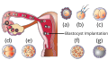
The above figure shows the various stages of development of a zygote as it passes through the oviduct and uterus: 

(a) The zygote divides into a two-celled stage via division or 1st cleavage while the zygote is still in the isthmus of the oviduct.

(b) Several more mitotic divisions or cleavages occur in the 2 celled stage to form a 2, 4, 8 and finally a 16-celled stage of the zygote. The 16 celled stage is called the morula and various cells formed after cleavage are called blastomeres.

(d) Morula then changes into a blastocyst after a few more divisions and this stage contains a fluid filled cavity in the embryo. The blastomeres become arranged and line up into an outer layer of cells called the trophoblast and an inner mass of cells. The fluid filled cavity is called blastocoel.

(e) Implantation of the embryo occurs at this blastocyst stage by the help of trophoblast layer which embeds itself into the uterine endometrium.


Q4Written answer

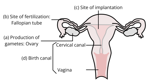
Draw a neat diagram of the female reproductive system and label the parts associated with the following (a) production of gamete, (b) site of fertilization (c) site of implantation and, (d) birth canal.

Show solutionHide solution
Worked solution

Ans:

seo images


a) Production of gamete is done in the ovaries lying on either side of the uterus. At one time, only one ovary releases the gamete.

b) Site of fertilization is the ampulla-isthmus junction in the oviduct or the fallopian tube of a human female.

c) Site of implantation in a human female is the uterine endometrium. However, ectopic pregnancies are fairly common where zygote gets implanted on the wrong site.

d) Birth canal is the cervical plus vaginal canal of a female i.e., the canal through which a grown fetus passes during its birth to come out of the womb.


Q5Written answer

With a suitable diagram, describe the organization of the mammary gland.

Show solutionHide solution
Worked solution

Ans: The mammary glands are paired structures meant mainly for the production of milk or newborn in a female after pregnancy has been achieved. These structures contain variable amounts of glandular tissue and huge amounts of fat. The glandular tissue of each breast is divided into about 15-10 mammary lobules which contain a group of cells called alveoli, the milk secreting cells. The milk secreted or made by these alveoli is stored in lumen of the alveoli until its transfer is required. This milk by the alveoli is transferred next to the mammary ducts into which the lumen or cavities of alveoli directly open. Many of these mammary ducts join together to form a lactiferous duct which opens up on the nipple or the areola. It is this opening through which a newborn sucks the milk out.

seo images